Chromosome Diagrams 101 Diagrams
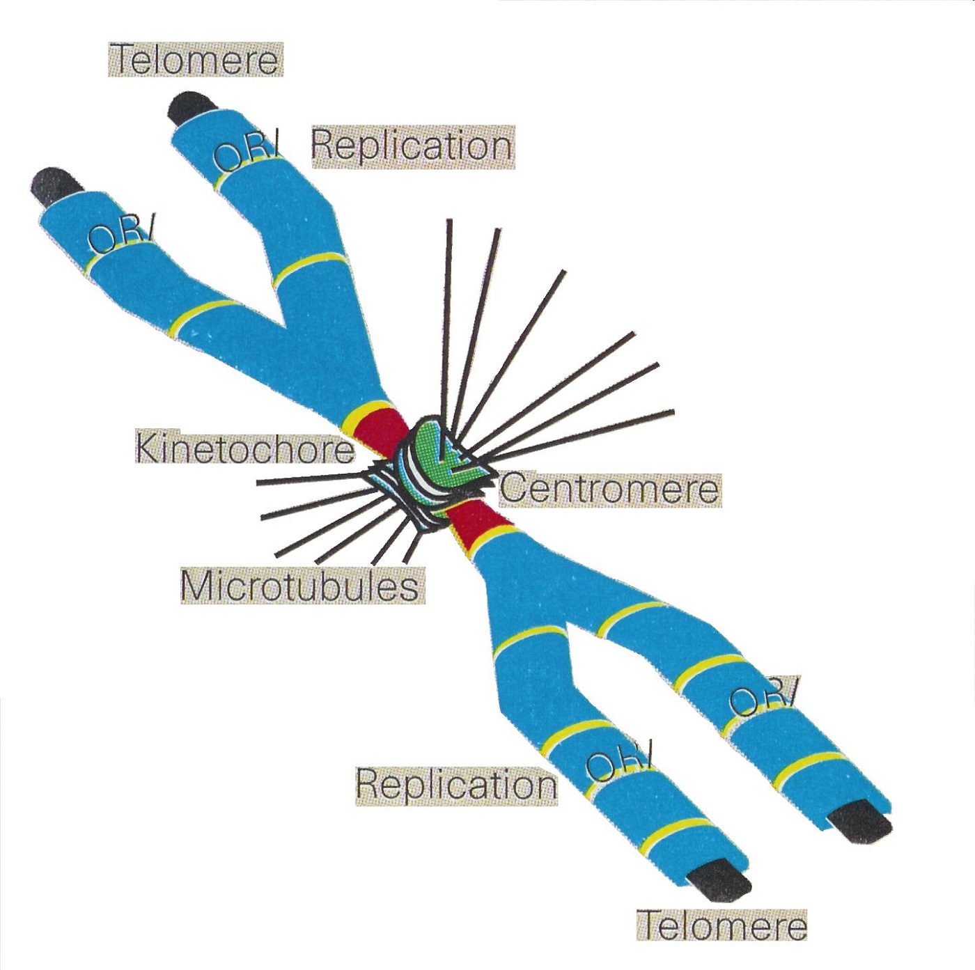
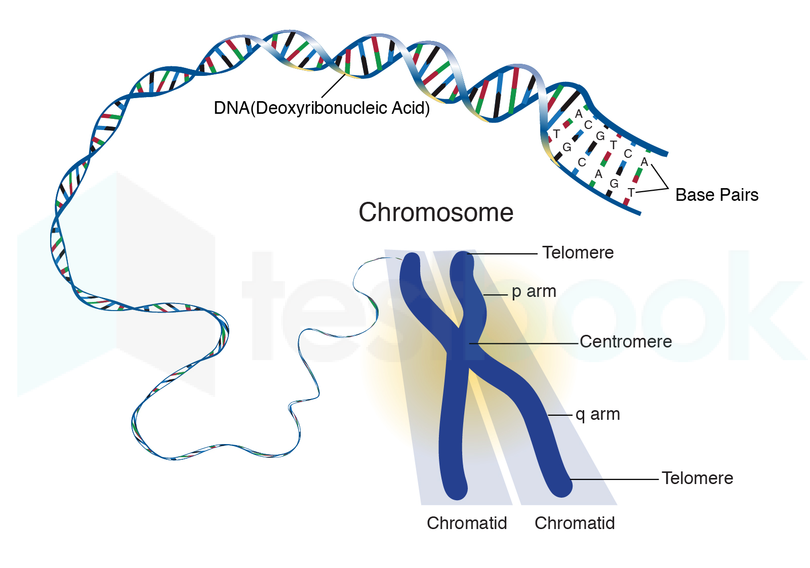
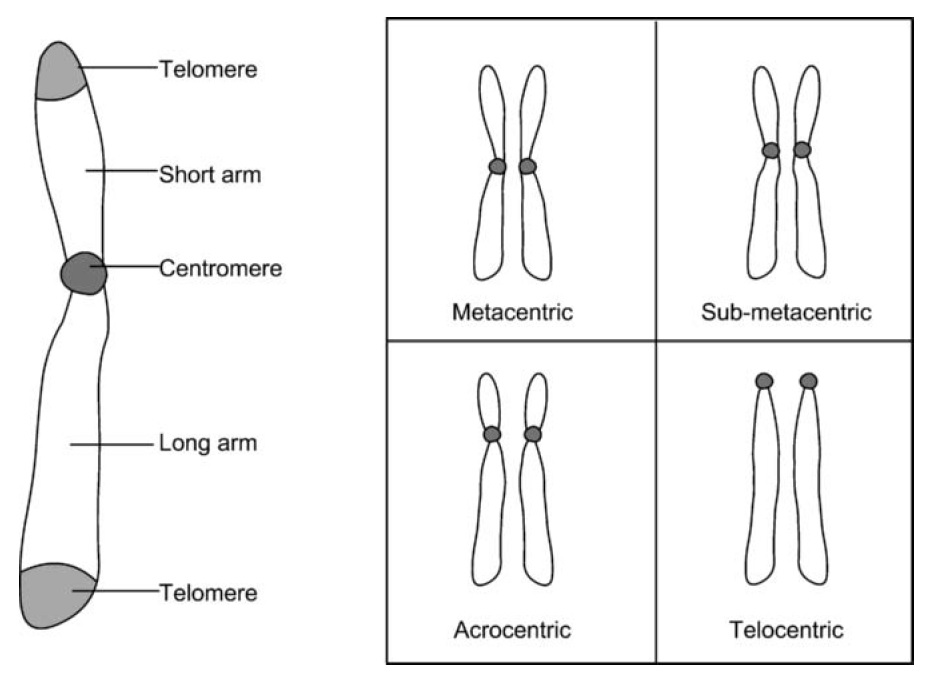
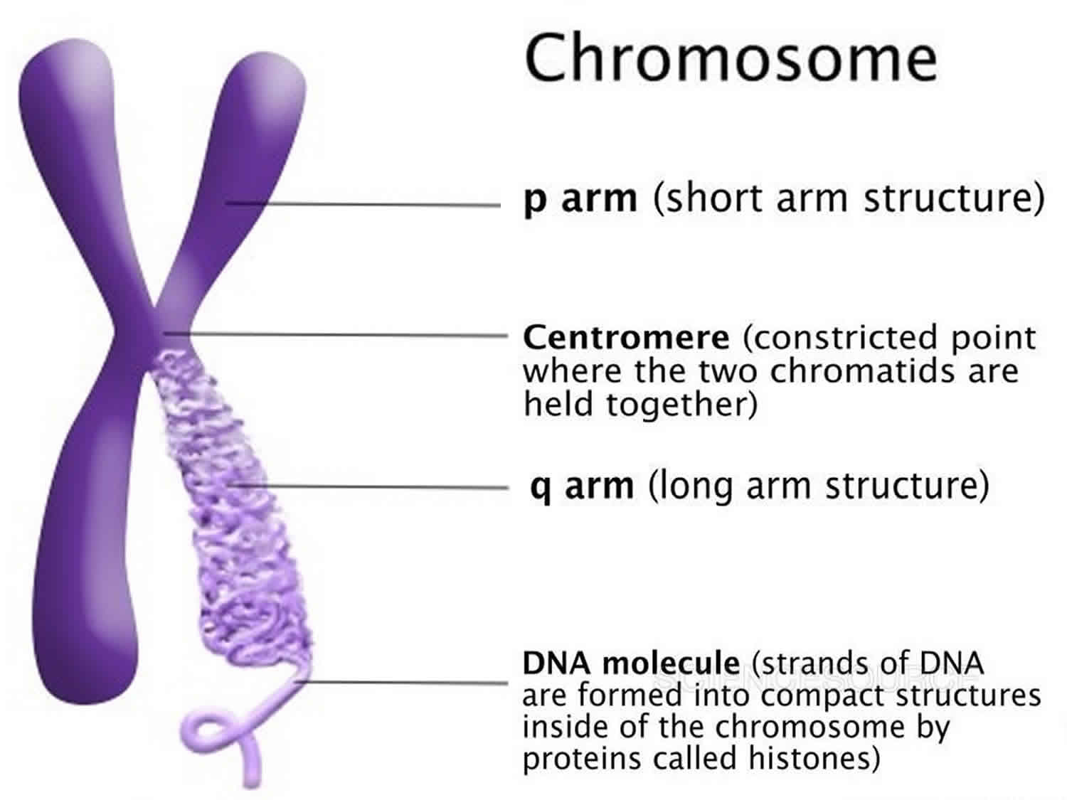
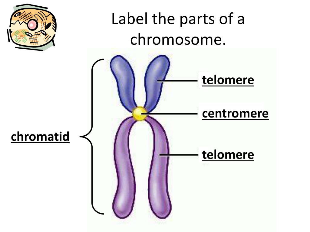
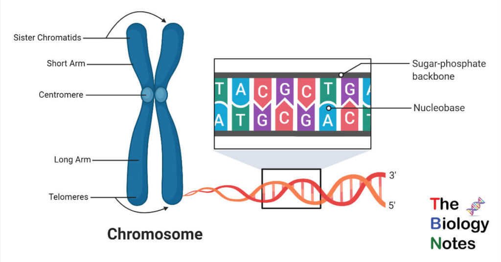
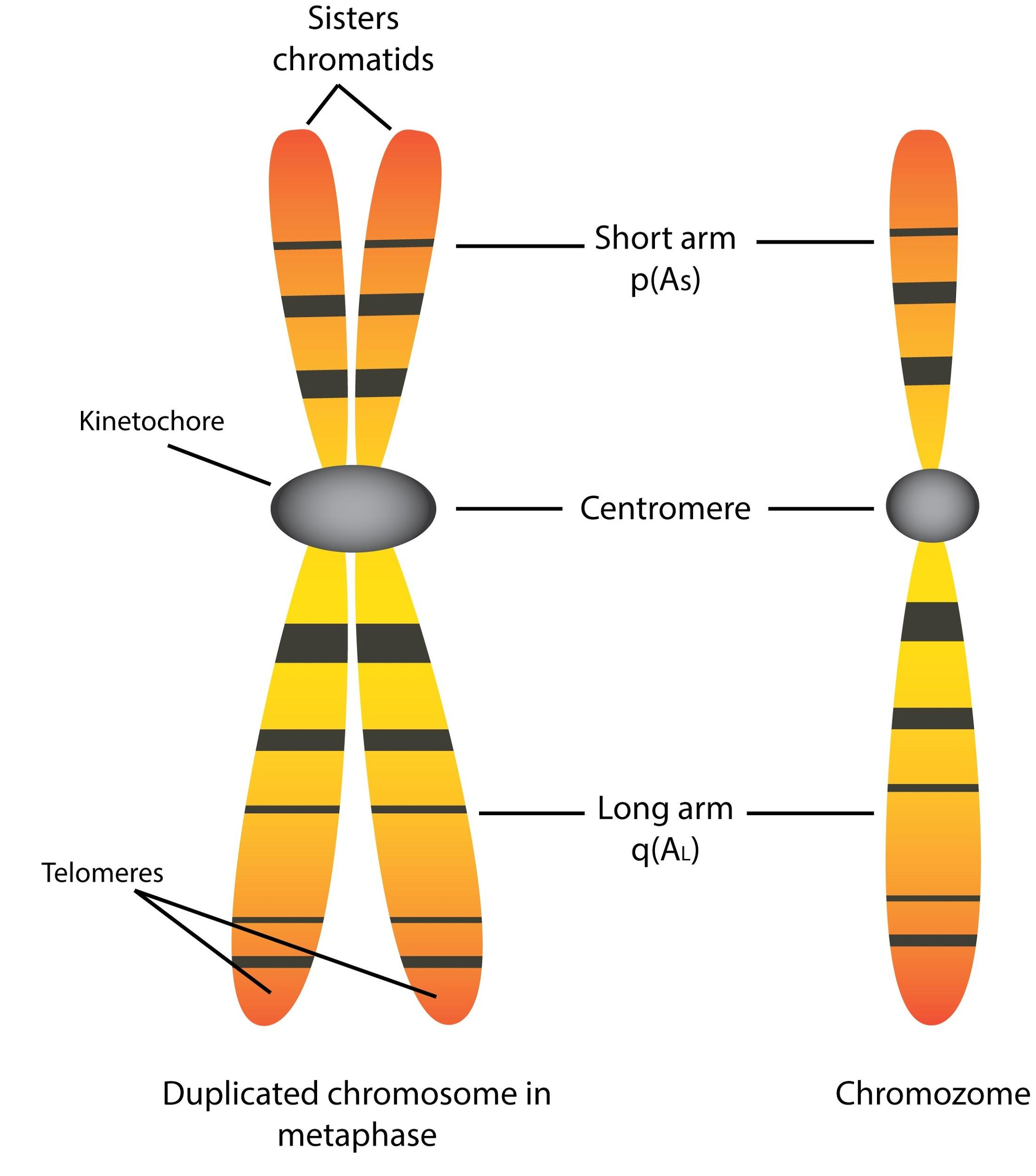
The structure of chromosomes. Each chromosome has a constriction point called the centromere, which divides the chromosome into two sections, or "arms." The short arm of the chromosome is labeled the "p arm." The long arm of the chromosome is labeled the "q arm." The ends of the chromosome are called telomeres.
[Solved] Number of chromosomes in human cell is
< Prev Next > Chromosome Map Our genetic information is stored in 23 pairs of chromosomes that vary widely in size and shape. Chromosome 1 is the largest and is over three times bigger than chromosome 22. The 23rd pair of chromosomes are two special chromosomes, X and Y, that determine our sex.
Structure and types of the eukaryotic chromosomes WikiLectures
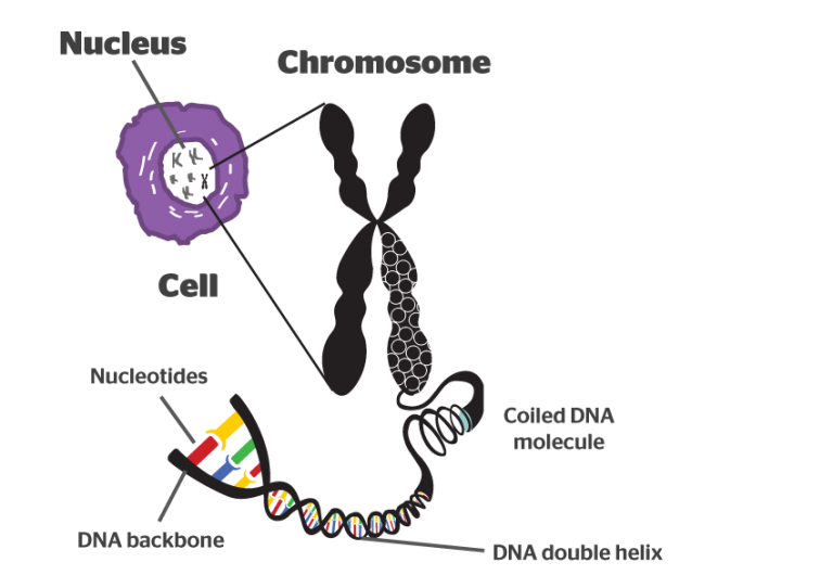
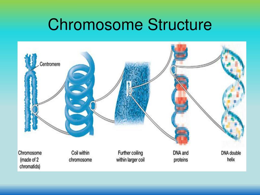
Genetics → Help Me Understand Genetics → Cells and DNA → What is a chromosome? What is a chromosome? In the nucleus of each cell, the DNA molecule is packaged into thread-like structures called chromosomes. Each chromosome is made up of DNA tightly coiled many times around proteins called histones that support its structure.
.PNG)
Cell Reproduction. Mitosis and Binary Fission Presentation Biology
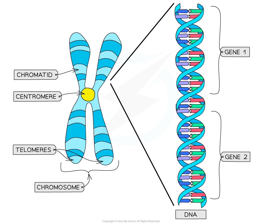
Chromosome Structure Labeling. Chromatid Chromosomes DNA Centromere Cell_Membrane Nucleus. This work is licensed under a Creative Commons Attribution-NonCommercial-ShareAlike 4.0 International License. Students label a simple diagram of a chromosome showing the centromere, chromatid, DNA, and the location of the chromosome within the nucleus of.
What is a Chromosome Definition, Structure and Evolution
DNA structure and function Google Classroom DNA is the information molecule. It stores instructions for making other large molecules, called proteins. These instructions are stored inside each of your cells, distributed among 46 long structures called chromosomes. These chromosomes are made up of thousands of shorter segments of DNA, called genes.
Chromosomes Introduction, Structure & Types A Level Biology Notes
Chromosome Definition. A chromosome is a string of DNA wrapped around associated proteins that give the connected nucleic acid bases a structure. During interphase of the cell cycle, the chromosome exists in a loose structure, so proteins can be translated from the DNA and the DNA can be replicated. During mitosis and meiosis, the chromosome.

Basic components of chromosome Telomeres, Cell Division, Chromosome, Dna, Peace
Posted June 3, 2019 in Cell Biology, Genetics, Worksheets by Shannan Muskopf centromere, chromatid, chromosome, DNA, label, nucleus, practice, structure A diagram of a chromosomein the nucleus of the cell. Students label the chromatid, centromere, chromosomes, cell membrane, DNA, and nucleus.

Biotechnology Basics of Cell, Nucleus, Chromosomes, DNA, RNA, Genes, Codons, Amino acids, etc
Chromosomes can be analyzed from living tissue and arranged in a karyotype (figure 13.1). Chromosomes can be sorted into the autosomal pairs (twenty-two) and sex chromosomes and classified to determine any abnormalities. A normal karyotype for a female is 46,XX, and a male is 46,XY. Deviations from this patterning can result in chromosomal.
CIE A Level Biology复习笔记5.1.1 Chromosome Structure翰林国际教育
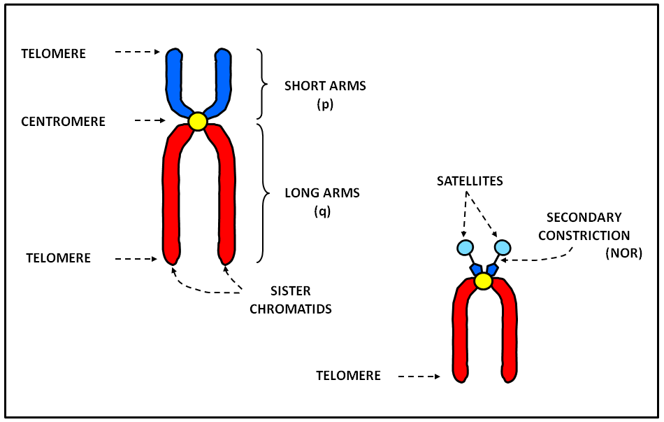
What are Chromosomes? Structure of a Chromosome Pellicle Matrix Chromonemata Centromere Secondary Constriction or Nucleolar Organiser Telomeres Types of Chromosomes A. Autosomes and Sex Chromosomes B. On the Basis of Number of Centromeres C. On the Basis of Location of Centromere Prokaryotic Chromosomes Eukaryotic Chromosomes a. Nucleosomes
PPT Chapter 8 Cell Reproduction PowerPoint Presentation ID322945
sohaib. 11 years ago. Chromosomes:A threadlike structure of nucleic acids and protein found in the nucleus of most living cells, carrying genetic information in the form of genes. Chromatid:Each of the two threadlike strands into which a chromosome divides longitudinally during cell division.
Karyotype, karyotype test & analysis, normal karyotype & abnormal karyotype
Google Classroom DNA, chromosomes, and genomes. Homologous chromosomes, sister chromatids, and haploid/diploid. Introduction When a cell divides, one of its main jobs is to make sure that each of the two new cells gets a full, perfect copy of genetic material.

Chromosome Structure, Illustration Stock Image C027/6970 Science Photo Library
Eukaryotic Chromosome Structure. Chromosome * s are long strands of DNA * in cells that carry genetic information. Most prokaryotic cells contain a single circular chromosome. Eukaryotic cells, with their much larger genomes, have multiple, linear chromosomes. The length and linear nature of eukaryotic chromosomes increase the challenge of.
31 Label The Parts Of The Chromosome Labels Design Ideas 2020
Chromosomes are thread-like structures located inside the nucleus of animal and plant cells. Each chromosome is made of protein and a single molecule of deoxyribonucleic acid (DNA). Passed from parents to offspring, DNA contains the specific instructions that make each type of living creature unique.
Chromosome History, Cell Division, Mutation, Disorders
Figure 1. Double-stranded DNA wraps around histone proteins to form nucleosomes that have the appearance of "beads on a string.". The nucleosomes are coiled into a 30-nm chromatin fiber. When a cell undergoes mitosis, the chromosomes condense even further. DNA replicates in the S phase of interphase.
Chromatid is(a) One half of chromosome(b) Haploid chromosome(c) Complete chromosome(d) Duplicate
This page titled 8.3: DNA Structure is shared under a CC BY license and was authored, remixed, and/or curated by Gerald Bergtrom. By 1878, a substance in the pus of wounded soldiers derived from cell nuclei (called nuclein) was shown to be composed of 5 bases (the familiar ones of DNA and RNA). The four bases known to make up..

Chromosome structure Chromosome, Chromosome structure, Structural biology
The compactness of chromosomes plays an important role in helping to organize genetic material during cell division and enabling it to fit inside structures such as the nucleus of a cell, the average diameter of which is about 5 to 10 μm (1 μm = 0.00l mm, or 0.000039 inch), or the polygonal head of a virus particle, which may be in the range of.